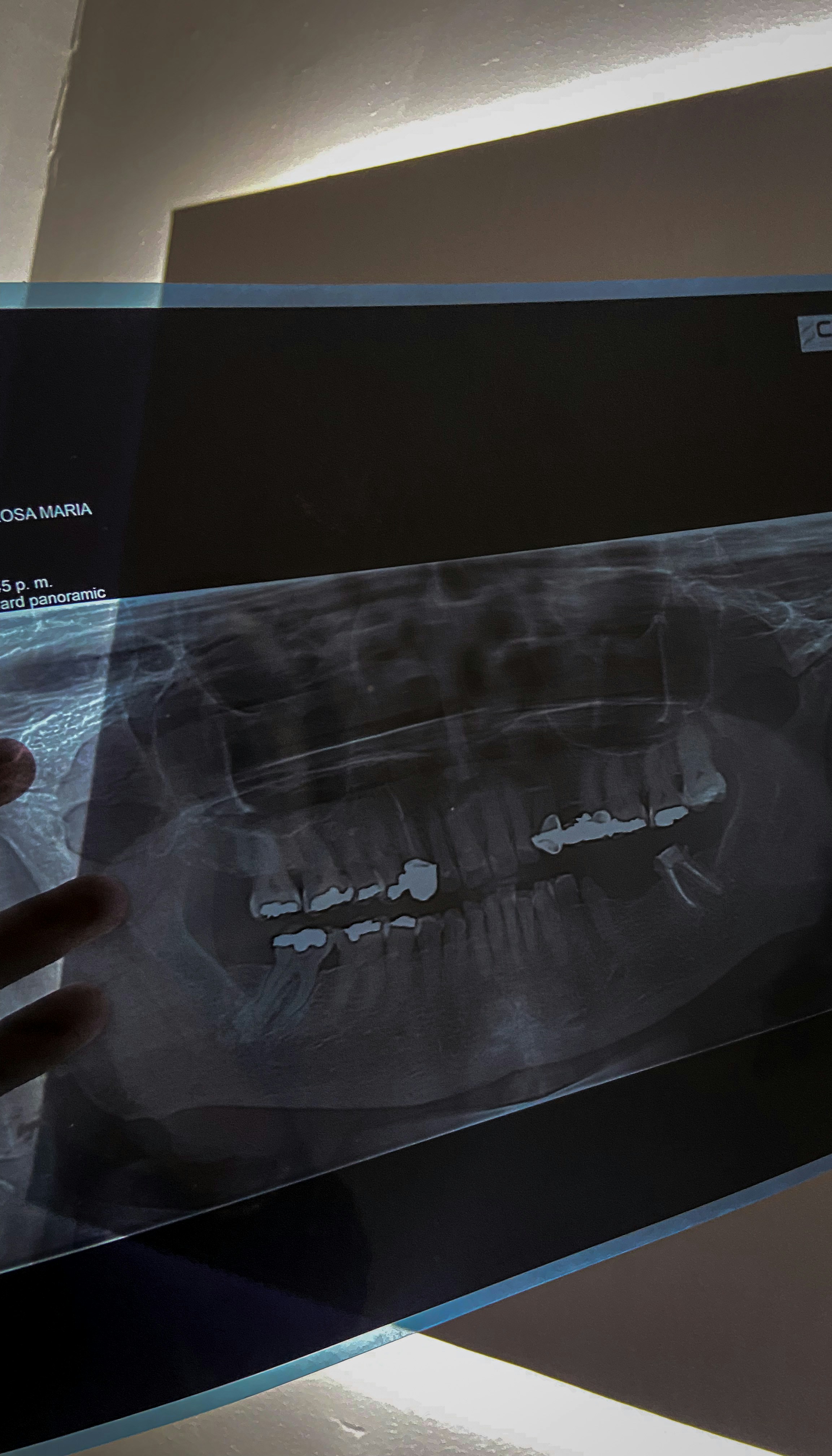
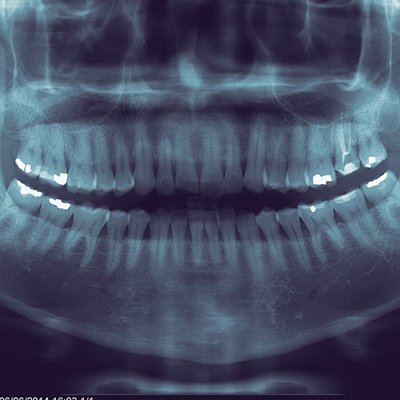
What is a Panoramic X-ray?
A panoramic X-ray is a specialized imaging technique used in dentistry to capture a comprehensive view of the entire dental arch in a single image. Unlike traditional dental radiographs, which typically focus on individual teeth or specific areas, a panoramic X-ray provides a 360-degree view, encompassing the upper and lower jaws, surrounding tissues, and structures such as the nasal cavity and sinuses. This wide-angle image is highly valuable for dental practitioners as it serves multiple purposes.
The panoramic X-ray process involves a rotating arm that moves around the patient’s head while the imaging receptor captures multiple images. These images are then digitally combined to create a seamless representation of the patient’s mouth and jaw. Due to this unique feature, panoramic X-rays are instrumental in identifying a variety of oral health concerns.
In particular, panoramic X-rays are crucial in orthodontics and oral surgery, as they allow professionals to assess the alignment of teeth, the position of wisdom teeth, and to evaluate the overall bone structure. Furthermore, this imaging technique aids in diagnosing various conditions such as cysts, tumors, and jaw disorders, which might not be easily detectable with standard intraoral X-rays.
Moreover, panoramic X-rays offer an advantage in terms of patient comfort, as the process does not require multiple images to be taken from different angles. Patients are generally seated in a stable position, reducing the discomfort that may arise from repositioning during traditional X-ray sessions. This comprehensive view, combined with increased patient comfort, makes panoramic X-rays an essential tool in modern dental practice.
How Does a Panoramic X-ray Work?
Panoramic X-rays, also known as panorex images, are an essential tool in dental diagnostics, offering a broad view of both the dental and skeletal structures. The technique employs a specialized X-ray machine that operates through a rather innovative process. The machine consists of a rotating arm that houses the X-ray source and a film or digital receptor positioned on the opposite side. When the X-ray procedure begins, the arm rotates around the patient’s head in a smooth motion, capturing a complete view of the anatomy within just a few seconds.
The functioning of the panoramic X-ray machine is based on a principle known as tomographic imaging. As the X-ray source moves, it emits a focused beam that traverses the area being imaged, passing through various tissues. The ability to capture images at a consistent and predetermined angle minimizes distortion, allowing for clear visualization of the jawbone, teeth, and surrounding structures. This controlled motion is pivotal, as it ensures that the images captured are of a high quality and represent a comprehensive anatomical layout.
Once the image is taken, the newly captured data is processed either digitally or via traditional film development techniques. In modern practices, digital receptors have largely replaced conventional films, streamlining the image acquisition and processing times. The resulting images are then enhanced using various software tools that allow dental professionals to analyze the structures with precision. These advanced technologies facilitate a clear depiction of jaw joint conditions, tooth alignment, and possible dental issues that may not be visible through standard radiographs. Thus, the mechanism of panoramic X-ray imaging not only ensures effective diagnostic capabilities but also enhances patient care through precise treatment planning.
Benefits of Panoramic X-rays
Panoramic X-rays, also known as orthopantomograms, provide a comprehensive view of the entire oral cavity, including the teeth, jawbone, and surrounding structures, in one single image. This characteristic is one of the primary advantages of panoramic X-rays, as they efficiently capture a wider spatial range compared to traditional intraoral X-rays, allowing dental professionals to assess the patient’s dental health thoroughly.
One significant benefit of panoramic X-rays is their ability to assist in the diagnosis of various dental and health issues. These images are invaluable in examining the position of wisdom teeth, detecting fractures, evaluating cysts or tumors, and assessing the condition of the jawbone. Additionally, they can be crucial for orthodontic treatment planning, as they reveal the relationship between teeth and facial structures. This capability enables clinicians to make informed decisions regarding treatment options.
Furthermore, panoramic X-rays expose patients to lower levels of radiation than traditional dental X-rays. While all radiographic procedures carry some risk due to radiation exposure, panoramic X-rays use advanced technology to minimize this risk, making them a safer choice for patients. The reduced radiation is particularly beneficial for children and those requiring multiple dental assessments, as it enhances patient safety while still providing vital diagnostic information.
In summary, panoramic X-rays are a crucial tool in modern dentistry, offering a wide range of diagnostic capabilities, reducing radiation exposure, and providing a broad view of dental structures. Their role in enhancing patient care cannot be overstated, making them a preferred choice among dental practitioners.
When Are Panoramic X-rays Used?
Panoramic X-rays serve as a vital diagnostic tool in various dental scenarios, helping practitioners capture a broad view of the patient’s oral structures. One of the primary situations where panoramic X-rays are employed is in orthodontics. These X-rays provide detailed images of the teeth and jaws, allowing orthodontists to assess the alignment of teeth and determine the best course of action for braces or other corrective measures.
Moreover, panoramic X-rays play a crucial role in dental implant planning. Before an implant procedure, clinicians use these X-rays to evaluate the bone structure and ensure adequate bone density and volume. This comprehensive view aids in the determination of the optimal implant size and location, thereby enhancing the likelihood of a successful implantation.
In addition to orthodontic evaluation and implant planning, panoramic X-rays are instrumental in assessing various jaw disorders. Conditions such as temporomandibular joint (TMJ) disorders, cysts, and tumors can be effectively diagnosed using this imaging technique. By providing an overview of the jaw’s anatomy, panoramic X-rays assist in formulating appropriate treatment plans tailored to the specific conditions identified.
Furthermore, panoramic X-rays are commonly utilized during routine screenings for oral health. Dentists recommend this imaging technique to detect potential dental issues early on, such as impacted teeth or bone loss. Unlike traditional X-rays, which focus on specific areas, panoramic X-rays provide a comprehensive snapshot of the oral cavity, making them a valuable tool for assessing overall oral health.
In conclusion, the application of panoramic X-rays is extensive, encompassing areas such as orthodontics, dental implant planning, and the evaluation of jaw disorders. Their role in routine oral health screenings further underscores their importance in modern dentistry, emphasizing the need for practitioners to incorporate this imaging technique into regular dental assessments.
Preparing for a Panoramic X-ray
Preparing for a panoramic X-ray is a crucial step that can contribute to the accuracy and efficiency of the imaging process. While generally straightforward, patients should adhere to several important guidelines to ensure optimal conditions for the examination.
First and foremost, clothing choices are significant. It is recommended that patients wear loose-fitting attire without any metallic elements. Items such as necklaces, earrings, and other jewelry should be removed prior to the X-ray, as they can interfere with the imaging results. In many facilities, patients are often required to wear a provided gown, which helps to eliminate any potential obstructions in the imaging area.
Dietary restrictions typically play a minimal role in preparing for a panoramic X-ray; however, it is advisable for patients to avoid consuming food or drinks containing metals shortly before the procedure. For instance, chewing gum or eating certain candy items can leave a residue that might affect the quality of the X-ray. Staying well-hydrated is encouraged, but patients should consult their healthcare providers regarding specific challenges that might arise based on individual health conditions.
Lastly, patients should ensure that they bring any necessary paperwork, including identification and insurance information, to their appointment. Consent forms may also be required, as they confirm that patients understand the procedure and its benefits and risks. If previously conducted X-rays or dental records are available, bringing these documents can also facilitate a comprehensive review by healthcare professionals. Overall, by following these preparation steps, patients can help ensure their panoramic X-ray experience is smooth and effective.
Potential Risks and Limitations
Panoramic X-rays are widely used in dental practices for their ability to provide a comprehensive overview of the oral and maxillofacial regions in a single image. However, like any medical imaging technique, they are not without potential risks and limitations that need to be acknowledged. One of the primary concerns is the exposure to radiation. While the amount of radiation emitted by panoramic X-ray machines is relatively low, it still poses a risk, particularly for vulnerable populations such as children and pregnant women. The long-term effects of cumulative radiation exposure are not fully understood, which necessitates careful consideration before proceeding with this imaging technique.
Another significant limitation associated with panoramic X-rays is their image quality, which can be compromised in certain cases. For instance, patients who have specific anatomical features, such as severe jaw deformities or those with multiple dental implants, may not receive clear and diagnostic images. The quality of the X-ray can vary based on the patient’s positioning and the technician’s skill, potentially leading to misinterpretations. Moreover, overlapping structures in the image might obscure critical details, making it challenging for dental professionals to accurately assess an individual’s oral health.
It is crucial for both practitioners and patients to weigh the benefits of panoramic X-rays against the potential risks and limitations. A thorough assessment of each patient’s unique anatomy and medical history can guide the decision-making process. Additionally, exploring alternative imaging modalities may be warranted for individuals who are at a higher risk or who may not benefit significantly from panoramic radiography. By being aware of these factors, practitioners can make more informed choices that prioritize patient safety and diagnostic accuracy.
Interpreting Panoramic X-ray Images
Interpreting panoramic X-ray images is a crucial skill for dental professionals, as these images provide a comprehensive view of the patient’s oral cavity, including jaws, teeth, and surrounding structures. Dental practitioners focus on several key factors when analyzing these two-dimensional images, which are invaluable for diagnosis and treatment planning.
One of the primary areas of focus is the assessment of tooth alignment and positioning. Dentists look for impacted teeth, such as wisdom teeth, that can cause overcrowding or alignment issues. Additionally, the radiographic image allows the dentist to evaluate the status of existing restorations and determine if they are functional or require replacement or further treatment.
Another critical aspect that dentists examine in panoramic X-rays is the presence of dental abnormalities. Common findings include cysts, tumors, or signs of infection such as periapical radiolucencies. These abnormalities can indicate various health conditions and will require appropriate follow-up actions, whether it’s monitoring or surgical intervention. Additionally, panoramic imaging aids in identifying periodontal disease by showing the level of bone surrounding the teeth, which is crucial for determining the health of the supporting structures.
Moreover, interpreting these images relies heavily on the dentist’s specialized training in radiology. Understanding the normal anatomy of the jaw and surrounding structures is vital for recognizing deviations from the norm. Advanced education and continuous training ensure that dental professionals remain up-to-date with the latest techniques and technology, enabling them to provide accurate diagnoses based on X-ray findings.
In summary, the interpretation of panoramic X-ray images is an essential component of modern dentistry. By analyzing these images meticulously, dentists can not only identify existing dental issues but also prevent future complications, ultimately contributing to improved patient outcomes.
Comparing Panoramic X-rays to Other Imaging Techniques
When considering various dental imaging techniques, it is essential to understand the distinctive features and applications of panoramic X-rays in comparison to other methods, such as bitewing images, periapical X-rays, and cone-beam computed tomography (CBCT). Each technique serves a unique purpose in dental diagnostics and treatment planning, facilitating comprehensive oral health assessments.
Panoramic X-rays, also known as orthopantomographs, provide a broad view of the entire mouth, showcasing the upper and lower jaws, teeth, and surrounding structures in a single image. This holistic view is particularly beneficial for orthodontic evaluations, wisdom tooth extractions, and identifying jaw abnormalities. In contrast, bitewing X-rays focus on the crowns of the upper and lower teeth. They are primarily utilized to detect interproximal caries and assess the health of the supporting bone, making them essential in routine dental check-ups.
Periapical X-rays, on the other hand, capture detailed images of a specific tooth or a small group of teeth from the root to the crown. This imaging technique is invaluable for diagnosing dental pain, abscesses, or detecting changes in the bone surrounding the tooth roots. While panoramic X-rays provide a comprehensive overview, they may lack the detailed focus necessary for evaluating individual teeth.
Lastly, cone-beam computed tomography (CBCT) represents a more advanced imaging technique that offers three-dimensional (3D) visualization of dental structures. CBCT is particularly useful for complex cases, such as implant planning or assessing jaw pathology, but it involves higher radiation exposure compared to traditional X-rays. In general, when choosing between these imaging techniques, a dentist evaluates the specific diagnostic requirements and the area of concern, ensuring the most appropriate method is employed for optimal results.
Future Trends in Panoramic X-ray Technology
The landscape of panoramic X-ray technology is evolving rapidly, driven by advancements in imaging methods and the integration of digital solutions. One significant trend includes the shift from traditional film-based X-rays to digital X-ray systems. Digital panoramic X-rays offer numerous advantages, such as reduced radiation exposure and immediate image acquisition, allowing for quicker diagnosis and treatment planning. Furthermore, digital formats enhance image quality, providing clearer and more detailed visuals compared to their film predecessors.
Another prominent development in the field is the application of artificial intelligence (AI) in image interpretation. AI technologies are being designed to assist dental professionals in diagnosing conditions from panoramic images. Machine learning algorithms can analyze patterns and abnormalities within the imaging data, improving accuracy and efficiency. As these systems become more sophisticated, they have the potential to support clinicians in making informed decisions, ultimately leading to better patient outcomes. AI can also streamline workflow by automating time-consuming tasks, such as measuring anatomical structures or identifying dental issues.
Moreover, enhancements in imaging techniques, including three-dimensional X-ray imaging, are set to revolutionize panoramic radiography. These advanced technologies allow for a multi-faceted view of the dental anatomy, facilitating more comprehensive assessments. By integrating 3D imaging with traditional panoramic views, practitioners can achieve a better understanding of complex cases, whether dealing with impacted teeth or assessing bone structure for implants. The combination of accessibility, precision, and reduced patient discomfort underscores the significant potential these innovations hold for the future of dental radiography.