Introduction to Femoral Condyles in Horses
The femoral condyles, which are crucial components of the knee joint in horses, consist of two distinct structures: the lateral femoral condyle and the medial femoral condyle. These bony prominences are situated at the distal end of the femur and articulate with the proximal tibia. The integrity and function of these condyles play a vital role in the overall biomechanics of the horse, particularly during locomotion.
The lateral femoral condyle is located on the outer side of the knee joint, while the medial femoral condyle is found on the inner side. Both condyles contribute to the stability and movement of the stifle joint, which is pivotal for activities such as walking, trotting, and galloping. This stability allows for smooth motion and effective weight transfer during different phases of ambulation.
Each condyle is critical in distributing loads and absorbs forces exerted on the knee during movement. Due to their weight-bearing nature, the lateral and medial femoral condyles are also susceptible to injuries and degenerative changes. Proper radiographic evaluation, particularly the lateral x-ray of horses, is essential for assessing any potential issues affecting these structures. Such insights can inform treatment and management strategies, ensuring the horse’s continued performance and well-being.
In equine radiology, understanding the differences between the lateral vs medial femoral condyle on lateral x-ray is fundamental. This knowledge equips veterinarians and equine practitioners with the ability to make accurate diagnoses, thereby facilitating targeted interventions. A comprehensive evaluation of both condyles on lateral x-ray enhances the ability to identify abnormalities, making early detection and treatment feasible.
The Importance of X-Rays in Equine Veterinary Medicine
In equine veterinary medicine, the utilization of X-ray imaging is fundamental for diagnosing injuries and conditions in horses. This non-invasive technique allows veterinarians to visualize internal structures, particularly the skeletal system, and is essential for identifying potential issues within a horse’s limbs. Among the various X-ray views, the lateral view provides critical insights into the anatomy of the femoral condyles, which are integral components of the horse’s stifle joint.
The lateral femoral condyle, located on the outer side of the femur, and the medial femoral condyle, found on the inner side, can both be assessed effectively using lateral X-rays of the horse. These two structures often face differing stresses and injuries during athletic activities, making it crucial to distinguish between them. The lateral view specifically enables veterinarians to evaluate the alignment, bone density, and presence of lesions or fractures in both the lateral and medial femoral condyle. Accurate assessment through lateral X-rays can reveal conditions such as osteochondritis dissecans, fractures, or signs of joint degeneration.
Furthermore, the importance of X-ray imaging extends beyond diagnosis; it is also vital for treatment planning. Understanding the specific issues affecting the lateral vs medial femoral condyle on lateral X-ray in horses enables veterinarians to devise the most effective treatment protocols and rehabilitation plans. Additionally, consistent monitoring through repeat X-rays can provide insights into the healing process, ensuring that any changes in the condition are detected promptly.
Anatomy of Lateral vs Medial Femoral Condyles
The femur, a critical bone in the horse’s hindlimb, features two distinct condyles at its distal end, specifically the lateral and medial femoral condyles. These condyles are essential for knee joint functionality and weight distribution. Understanding the anatomical differences between the lateral vs medial femoral condyle on lateral x-ray of a horse aids in diagnosing potential pathologies.
The lateral femoral condyle is typically larger and more rounded when compared to the medial condyle, which tends to be slightly smaller and more elongated. This shape difference plays a significant role in the biomechanics of the joint, providing stability and acting as a point of attachment for numerous ligaments and tendons. On a lateral x-ray, the lateral femoral condyle appears more prominent, making it easier to identify.
In terms of location, the lateral condyle is situated outside (laterally) of the midline, while the medial condyle lies toward the center of the horse’s body. This orientation is critical for resulting joint motion during flexion and extension. Notably, the condylar aspect of the femur is covered by cartilage, which is crucial for smooth articulation with the tibia. Abnormalities in the condylar structure or alignment can lead to clinical implications, including joint pain and reduced mobility.
Furthermore, varying weight distribution during movement may lead to different stress levels on the two condyles, making it essential to evaluate these structures adequately during veterinary examinations, particularly through lateral x-ray imaging. Understanding the anatomical and structural differences assists veterinarians in identifying conditions such as osteochondritis dissecans or fractures associated with the lateral vs medial femoral condyle on lateral x-ray in horses. This knowledge is invaluable for ensuring the health and performance of equine patients.
Interpretation of Lateral X-Rays
Understanding the interpretation of lateral X-rays is crucial for evaluating the femoral condyles in horses, specifically when comparing the lateral vs medial femoral condyle on lateral X-ray. These images provide valuable insights into bone morphology and the overall structure of the knee joint. Careful analysis can help veterinarians identify normal versus abnormal findings, which is essential for accurate diagnosis and treatment options.
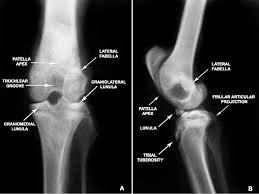
When examining lateral X-rays, it is important to focus on several key factors. First, the alignment of the femoral condyles should be considered. The normal anatomy of the femoral condyles shows a smooth contour, with the lateral condyle typically being slightly larger than the medial condyle. Any deviation from this standard may indicate pathological changes, such as osteoarthritis or fractures.
Next, special attention should be paid to the angles formed by the condyles and the surrounding bony structures. The patellar and femoral alignment can help determine whether there is any joint incongruity, which may signify an underlying issue. In addition, the presence of any radiolucent lesions or irregularities along the bony surfaces can provide further clues to potential condylar damage.
It is also essential to assess soft tissue structures surrounding the femoral condyles. Swelling, fluid accumulation, or abnormal opacities around the joint may indicate injury or disease. By comparing both the lateral and medial femoral condyle on lateral X-ray images, practitioners can develop a comprehensive view of any issues present and formulate appropriate treatment strategies.
Overall, a meticulous approach to interpreting lateral X-rays ensures that clinicians are well-equipped to identify variations in the femoral condyles. This knowledge culminates in improved veterinary care and better outcomes for the equine patient.
Common Injuries and Conditions Associated with Femoral Condyles
The femoral condyles in horses, specifically the lateral and medial components, play a crucial role in the overall function of the knee joint. Various injuries and conditions can affect these structures, leading to significant clinical implications for equine athletes. Common injuries include fractures, which typically result from trauma or excessive stress. Fractures can occur in either the lateral or medial femoral condyle, and lateral X-rays are pivotal in diagnosing such conditions. On these X-rays, fractures may appear as discontinuities or irregularities in the bone architecture, requiring careful evaluation by a veterinarian.
Another prevalent condition affecting the femoral condyles is osteochondritis dissecans (OCD). This developmental disorder is characterized by the improper formation of cartilage and underlying bone, often resulting in joint pain and lameness. The condition frequently impacts the lateral femoral condyle, with lateral X-rays revealing irregularities or lesions indicative of OCD. Recognition of these changes is crucial for prompt treatment, which may involve both medical and surgical interventions to mitigate the horse’s discomfort and restore optimal function.
Furthermore, arthritic changes are common conditions associated with the femoral condyles in horses, particularly in older or heavily worked animals. Osteoarthritis can lead to alterations in the joint surfaces, resulting in pain and diminished range of motion. On lateral X-rays, early signs may include the formation of osteophytes or joint effusion, both of which signify degenerative changes in the joint. A thorough understanding of these conditions and their radiographic appearances is integral to diagnosing and managing injuries involving the lateral vs medial femoral condyle on lateral X-ray in horses, ultimately improving outcomes and performance.
Clinical Significance of Observing Lateral vs Medial Differences
The evaluation of lateral vs medial femoral condyle on lateral x-ray in horses plays a crucial role in veterinary diagnostics and treatment planning. The femoral condyles are the rounded protrusions on the distal end of the femur, serving important functions in the knee joint. Observing these structures can unveil critical anatomical and pathological differences that influence both diagnosis and therapeutic approaches.
Understanding the lateral and medial femoral condyles is essential for diagnosing various conditions such as osteochondrosis, fractures, and articular cartilage wear. More specifically, lateral condyle injuries may manifest differently than those of the medial condyle, which can result in variations in symptoms, lameness, and overall joint functionality. Accurately identifying which condyle is affected can lead to more targeted and effective treatment strategies, thereby reducing the time to recovery and enhancing the horse’s long-term welfare.
Furthermore, recognizing the lateral vs medial discrepancies on x-ray allows practitioners to tailor rehabilitation programs and preemptively address potential complications. For instance, a lesion on the medial femoral condyle might require a different management approach than a similar one on the lateral side. When practitioners understand the importance of these distinctions, they can advocate for the most appropriate surgical interventions, medication, or physical therapy regimens suited to the specific needs of the horse.
The ability to discern between lateral and medial femoral condyles not only underscores the importance of precise imaging but also reinforces the necessity of a holistic approach in equine medicine. By considering these factors, veterinarians can significantly improve diagnosis precision, enhance treatment options, and ultimately lead to better outcomes for the horse.
Case Studies: X-ray Findings and Clinical Outcomes
Understanding the lateral vs medial femoral condyle on lateral x ray horse is crucial for accurate diagnoses and treatment planning in equine medicine. This section explores several case studies that highlight how specific findings on X-ray images can correlate with clinical outcomes.
In one case, a 7-year-old thoroughbred presented with lameness in the right forelimb. The lateral X-ray revealed significant irregularity in the lateral femoral condyle, indicative of osteochondritis dissecans (OCD). Due to this finding, the attending veterinarian recommended surgical intervention to address the fragmented cartilage. Post-operative follow-up indicated a marked improvement in lameness and increased performance in competitive events, demonstrating the effectiveness of timely surgical treatment following precise X-ray interpretation.
Another case involved a 10-year-old Quarter Horse that exhibited chronic joint pain in the left hind leg. Lateral X-rays showed subtle sclerosis of the medial femoral condyle, suggestive of early degenerative changes. The clinical decision was made to initiate a conservative management plan involving joint supplements and controlled exercise. After several months, subsequent X-rays indicated stabilization of the condition, and the horse reported improved mobility without surgery, showcasing the importance of monitoring and adjusting treatment based on X-ray findings.
These cases emphasize the critical role of lateral X-ray evaluations in identifying conditions specific to the femoral condyles. The findings not only guide immediate clinical decisions but also significantly influence long-term outcomes. Understanding the differences between the lateral and medial aspects of the femoral condyles through careful X-ray assessment is essential for effective equine care.
Preventative Measures and Care for Equine Health
Maintaining the health of a horse’s femoral condyles, particularly the lateral vs medial femoral condyle on lateral x-ray, requires a proactive approach that encompasses various aspects of the horse’s lifestyle and care. Equine health is influenced significantly by diet, exercise, and management practices, which all play vital roles in ensuring joint integrity.
Firstly, providing a balanced diet rich in essential nutrients is fundamental. Horses require adequate levels of vitamins, minerals, and proteins to support their muscle and bone health. Nutrients such as omega-3 fatty acids and antioxidants can help reduce inflammation in the joints, contributing positively to the health of the femoral condyles. Incorporating supplements specifically designed for joint health can also be beneficial; these often contain glucosamine and chondroitin which aid in cartilage maintenance.
Secondly, appropriate exercise is crucial. Regular, low-impact activities help strengthen the muscles surrounding the joints, providing necessary support and reducing the likelihood of injury. Overexertion, particularly on hard surfaces, can lead to stress on the medial and lateral femoral condyles, potentially resulting in damage detectable through lateral x-rays. Therefore, tailoring an exercise regimen that fits the horse’s age, breed, and condition is essential.
Furthermore, regular veterinary check-ups and joint evaluations are recommended as part of preventative care. Early detection of issues related to the femoral condyles can prompt more effective treatment and management before they escalate into severe problems. Ensuring that the horse has access to soft, clean bedding can also minimize the risk of joint trauma during rest periods.
Additionally, maintaining a healthy body weight is vital, as obesity can place excessive stress on the joints, exacerbating potential issues with the lateral vs medial femoral condyle on lateral x-ray in horses. By integrating these various strategies—nutrition, exercise, veterinary care, and weight management—horse owners can proactively safeguard their equine companions’ joint health and overall well-being.
Conclusion and Future Perspectives in Equine Imaging
In summarizing the important distinctions between the lateral and medial femoral condyle on lateral X-rays in horses, it is evident that accurate imaging plays an essential role in diagnosing and managing equine conditions, particularly those related to the stifle joint. Understanding the anatomical differences and potential pathological changes of these femoral condyles is crucial for equine veterinarians as it can significantly influence treatment choices and outcomes.
Future advancements in imaging technology hold great promise for enhancing our ability to evaluate the lateral vs medial femoral condyle on lateral X-ray in horses. Innovations such as improved imaging resolution and techniques, including digital radiography and potentially MRI or CT integration, may allow for a more comprehensive view of the joint structures. These advancements could lead to early detection of subtle changes that are often missed in standard X-ray views, thereby allowing for timely interventions.
Moreover, the incorporation of machine learning and artificial intelligence into imaging analysis may further improve diagnostic accuracy and efficiency. Such technologies can assist veterinary professionals in accurately interpreting images and may be particularly useful in distinguishing between normal anatomical variations and pathological conditions affecting the femoral condyles. Enhancing these capabilities will not only benefit our understanding of equine joint health but also improve the overall management strategies for these animals.
Ultimately, as imaging techniques continue to evolve, the veterinary community will gain a deeper understanding of conditions associated with the lateral vs medial femoral condyle, paving the way for improved health outcomes for horses. Continued research and development efforts are required to ensure that future imaging modalities remain at the forefront of equine medicine.