Introduction to Cavities and Fillings
Dental cavities, also known as caries or tooth decay, represent a common oral health issue that affects individuals of all ages. They are characterized by the gradual destruction of tooth structure, primarily caused by the interaction between acids produced by bacteria in the mouth and the minerals in the tooth enamel. When bacteria break down sugars and carbohydrates from food, they release acids that can demineralize the enamel, leading to the formation of cavities.
The genesis of cavities typically begins with plaque, a sticky film comprised of bacteria that adheres to the teeth. If not effectively removed through regular brushing and flossing, plaque can harden into tartar, further increasing the potential for decay. Factors contributing to the prevalence of cavities include poor oral hygiene practices, a diet high in sugars and carbohydrates, and individual genetic predispositions that may affect tooth resilience and enamel strength.
Notably, the importance of treating cavities with fillings should not be underestimated. When left untreated, cavities can lead to more severe dental issues, including infections, abscesses, or even tooth loss. Fillings serve as an effective means to restore both the function and integrity of the affected tooth. These restorative materials, which can include composite resins, metals, or glass ionomers, fill the cavity and protect the tooth from further decay and potential sensitivity.
In summary, understanding cavities, their formation, and the significance of fillings is essential for maintaining optimal oral health. Recognizing the risk factors and encouraging proactive dental care can help prevent the onset of cavities, ensuring a healthier smile for years to come.
Types of Filling Materials
Cavity fillings are essential in restoring the function and integrity of a tooth that has experienced decay. There are several materials used for this purpose, each with unique properties, advantages, and disadvantages. Here, we explore the most common types of filling materials: amalgam, composite, resin, glass ionomer, and gold.
Amalgam fillings are made from a blend of metals, primarily silver, mercury, tin, and copper. These fillings are well-known for their durability and strength, making them suitable for posterior teeth where chewing forces are significant. However, their metallic color can be a drawback for aesthetic concerns, particularly in visible areas.
Composite fillings consist of a resin material that is tooth-colored, allowing for a natural appearance. This option is favored for front teeth due to its aesthetic qualities. While composite fillings bond well to the tooth structure, they may not be as durable as amalgam, particularly in high-stress areas, which means they could require replacement sooner under heavy use.
Resin-based materials offer a modern solution and are particularly advantageous for smaller cavities. Similar to composites, they blend well with natural tooth color and can be polished to match the surrounding enamel. However, their wear resistance can be limited, and light-cured versions may require more time during the procedure to harden.
Glass ionomer fillings release fluoride, which can help in the prevention of further decay. They are suitable for areas that are not subjected to heavy chewing forces and are often used in pediatric dentistry. However, these fillings are more prone to wear and not as aesthetically pleasing as composite materials.
Gold fillings are highly durable and are appreciated for their long lifespan. They, however, come at a higher cost and are more noticeable than other options. In select situations, the benefits of gold fillings can outweigh the aesthetic concerns, making them a solid choice for posterior teeth.
The Role of X-Rays in Diagnosing Cavities
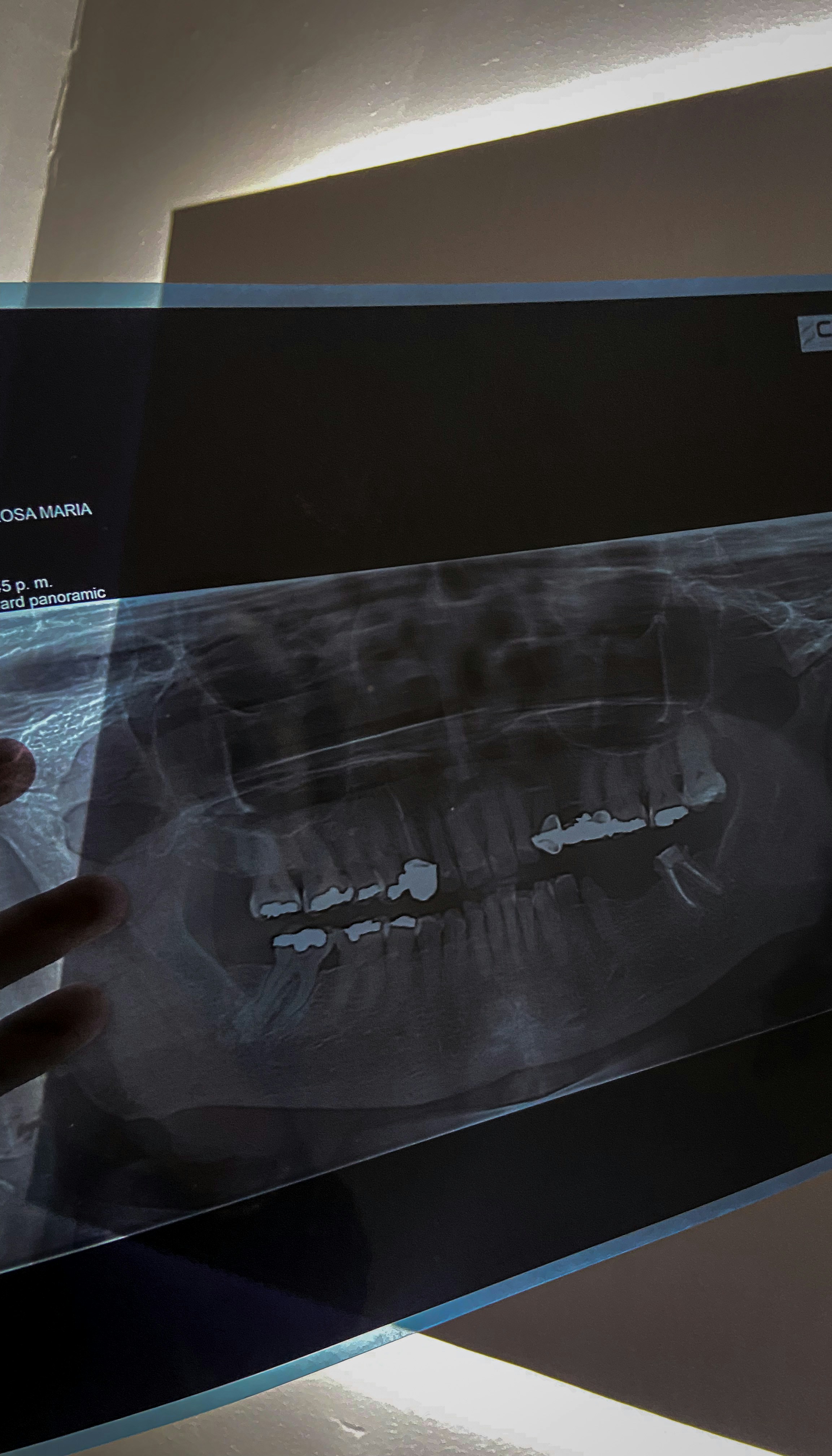
X-rays play a crucial role in the dental field, particularly in the diagnosis of cavities. These imaging tools allow dentists to visualize the internal structure of teeth and surrounding bone, helping them identify issues that may not be apparent during a routine examination. By using X-rays, dentists can detect cavities at their earliest stages, enabling more effective treatment and increased chances of preserving the tooth structure.
There are several types of dental X-rays employed for cavity detection. One of the most common is the bitewing X-ray, which captures images of the upper and lower teeth in a specific area of the mouth. This type of X-ray is particularly useful in identifying cavities between teeth as well as assessing the bone levels supporting the teeth. Bitewing X-rays are generally taken during routine checkups and can reveal hidden cavities effectively.
Another type is the periapical X-ray, which shows the entire tooth from its crown down to the bone that supports it. This type of imaging can uncover cavities that have progressed to affect the root of the tooth or surrounding structures. Additionally, periapical X-rays are invaluable in diagnosing conditions beyond cavities, such as abscesses or bone loss.
Furthermore, advancements in dental technology have introduced digital X-rays, which emit less radiation and produce high-quality images almost instantaneously. This quick turnaround not only aids in immediate diagnosis but also enhances patient comfort by reducing wait times. Ultimately, X-rays are indispensable tools for dentists, allowing for accurate diagnosis and timely intervention in the treatment of cavities, thereby safeguarding the overall oral health of patients.
How X-Rays Influence Filling Procedures
X-rays play a crucial role in modern dentistry, especially when it comes to diagnosing dental issues and planning appropriate treatments, such as cavity fillings. These imaging tools provide a detailed view of the teeth and surrounding structures, allowing dentists to assess the extent of decay more accurately. By revealing the internal condition of a tooth, X-rays can identify how deep the decay penetrates, which is essential for determining the most suitable type of filling needed.
Moreover, X-rays are instrumental in detecting multiple lesions that may not be readily visible during a standard dental examination. This capability ensures that all areas of concern are addressed, preventing further complications that could arise from untreated cavities. When multiple cavities are detected through X-ray imaging, dentists can formulate a comprehensive treatment plan that addresses all affected areas effectively, minimizing the number of separate visits a patient may need.
Additionally, X-ray images help in planning the filling procedure itself. Dentists utilize the information gleaned from these images to decide on the most effective treatment strategy, considering factors such as the material to be used for the filling and the technique required for optimal placement. For instance, knowing the precise location and extent of the decay allows the dentist to choose between direct or indirect filling methods accordingly. This tailored approach ensures that the filling is both effective and long-lasting, contributing to better oral health outcomes.
In conclusion, X-rays significantly enhance the cavity filling process by providing essential insights into the severity and nature of dental decay. Their role in assessing the dental landscape ensures that cavity fillings are not only reactive but also preemptive in nature, promoting long-term dental health.
The Procedure for Getting a Cavity Filled
When addressing a cavity, the first step is typically an initial consultation with a dental professional. During this appointment, the dentist will assess the patient’s dental health and recommend necessary treatments. This often includes taking dental X-rays, which provide detailed images of the teeth and help identify the extent of the cavity. X-rays are essential for accurate diagnosis, as they reveal underlying issues that may not be immediately visible during a visual examination.
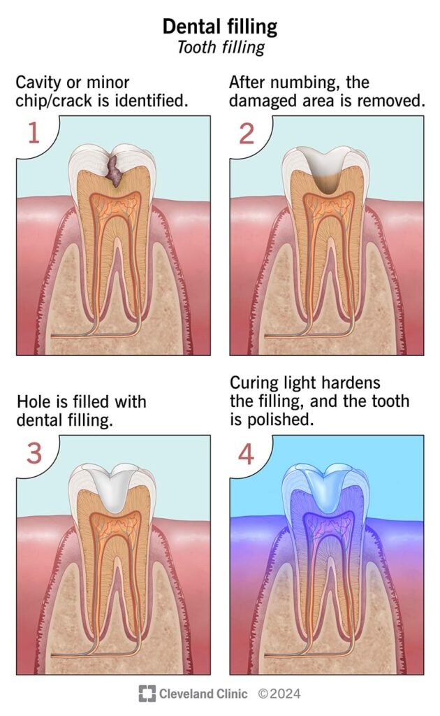
Once a cavity has been confirmed through X-ray analysis, the next step is administering anesthesia. The dentist applies a local anesthetic to numb the affected area, ensuring that the patient remains comfortable during the procedure. It is important for patients to communicate any allergic reactions to anesthesia to their dentist prior to this step. Once the anesthesia takes effect, the dentist will use specific tools to remove the decayed portion of the tooth.
After the decayed material is removed, the cavity is cleaned to eliminate any bacteria or debris, preparing it for the filling. Depending on the extent of the cavity and the material used, the filling process can vary. Common filling materials include composite resins, amalgam, and glass ionomer. The chosen filling material is inserted into the cavity and shaped to fit the tooth’s structure. The dentist may use a curing light to harden composite fillings, ensuring a secure bond.
Once the filling is placed, the dentist will check the bite to ensure proper alignment. After making any necessary adjustments, the dental team will provide post-treatment care instructions. It is crucial for patients to maintain good oral hygiene practices following the filling procedure and schedule regular dental checkups to monitor their dental health effectively.
Risks and Considerations of Fillings and X-Rays
When considering dental treatments such as cavity fillings and X-rays, it is essential to understand the potential risks and considerations involved. Dental fillings are often made from materials like amalgam, composite resin, or porcelain, and each of these substances carries its own risk of allergic reactions. Some individuals may be sensitive to specific components of the filling material, leading to adverse reactions such as inflammation or irritation in the affected area.
Alongside allergic reactions, patients may experience discomfort during and after the filling procedure. Local anesthesia is commonly used to minimize pain, yet some residual sensitivity can occur as the anesthesia wears off. It is important for patients to communicate with their dentists about any concerns regarding pain management and to follow post-treatment care instructions to alleviate discomfort.
Another critical aspect is the use of dental X-rays, which are frequently employed to diagnose and treat dental issues more effectively. While X-rays are a valuable diagnostic tool, they do expose patients to a small amount of radiation. Although reputable studies indicate that the risk of developing radiation-induced complications is minimal, this concern can be magnified in vulnerable populations, such as pregnant women and children. Dentists typically exercise caution and employ protective measures, such as lead aprons, during X-ray procedures to mitigate exposure risks.
Moreover, it is crucial to discuss the necessity of each X-ray with a healthcare professional, as the benefits of obtaining clear images for accurate diagnosis may outweigh the risks associated with radiation exposure. Ongoing advancements in dental technology aim to reduce radiation doses further while maintaining high diagnostic efficacy. Patients should feel empowered to ask questions and express concerns regarding their treatment options, ensuring that decisions made align with their individual health considerations.
Aftercare for Dental Fillings
Following a dental filling procedure, it is essential to adhere to recommended aftercare practices to ensure optimal healing and the longevity of the filling. One common experience after receiving a filling is sensitivity. It is advisable to manage this sensitivity by avoiding very hot or cold foods and beverages for at least 24 hours post-treatment. Over-the-counter pain relievers can help alleviate any discomfort, although it is important to consult with your dentist if sensitivity persists beyond a few days.
Another crucial aspect of post-filling care involves dietary considerations. Initially, it is recommended to stick to a soft food diet. Foods that are too hard or crunchy can put undue pressure on the newly filled tooth, potentially jeopardizing its integrity. Additionally, try to avoid sticky substances like caramel or chewing gum, as these can dislodge the material or create additional challenges for the filling.
Maintaining a well-rounded oral hygiene routine is vital after receiving a dental filling. Gentle brushing with a soft-bristled toothbrush will help keep the area around the filling clean without causing irritation. It is also advisable to use a fluoride toothpaste, which can strengthen the enamel and assist in preventing future cavities. Flossing daily is equally important, but care should be taken not to use excessive force around the filled area. If there are any concerns about how to floss adequately without disturbing the filling, discussing this with your dentist can provide tailored advice.
Regular dental check-ups remain essential, as these appointments allow the dentist to monitor the condition of the filling and ensure that oral health is well-maintained. If any problems arise, such as noticeable pain or changes in sensation, it is crucial to contact your dentist promptly for evaluation and necessary treatment.
FAQs about Cavities, Fillings, and X-Rays
When it comes to dental health, particularly concerning cavities, fillings, and the accompanying X-ray procedures, patients often have numerous questions. Addressing these common queries can greatly enhance understanding and alleviate concerns.
How often should I have X-rays taken? The frequency of dental X-rays primarily depends on individual health history and the dentist’s evaluation. For most individuals, X-rays are typically taken every one to two years. However, patients with a higher risk of cavities may require more frequent imaging. Regular check-ups and assessments by a dental professional will help determine the optimal schedule for X-rays.
Do fillings need to be replaced? Over time, dental fillings can deteriorate due to various factors such as wear and tear, recurrent decay, or changes in the surrounding tooth structure. It is essential to have regular dental check-ups to assess the condition of existing fillings. If a filling is cracked, loose, or shows signs of wear, the dentist will recommend replacing it to maintain oral health and prevent further complications.
What can I expect during and after the filling procedure? During a filling procedure, the dentist will first numb the affected area to ensure comfort. The decayed portion of the tooth is removed, and the filling material, which can be composite resin, amalgam, or other materials, is inserted and shaped to restore function. After the procedure, it is common to experience slight sensitivity, but this should subside in a few days. Patients are advised to follow post-operative care instructions provided by their dentist to support healing and manage any discomfort.
Understanding the answers to these frequently asked questions can empower patients to make informed decisions regarding their dental care, enhancing both their knowledge and confidence in managing oral health.
Conclusion and Final Thoughts
In this comprehensive guide on cavity fillings and X-rays, we have explored significant aspects of dental care that play a crucial role in preserving oral health. The detection and treatment of cavities are paramount to preventing more severe dental issues. Regular dental check-ups are essential in identifying cavities in their early stages. These visits allow dental professionals to assess, diagnose, and treat any tooth decay effectively, often preventing the need for more invasive procedures in the future.
X-rays serve as an invaluable tool in the detection of cavities that may not be visible during a standard oral examination. They provide insight into underlying problems, aiding dentists in developing tailored treatment plans. By enabling the identification of hidden cavities between teeth and beneath restorations, X-rays assist in maintaining overall dental health. Utilizing X-ray technology in conjunction with regular dental visits can dramatically improve the likelihood of timely interventions, ultimately reducing discomfort and costs associated with advanced decay.
Moreover, preventative strategies, such as maintaining proper oral hygiene practices and dietary awareness, contribute significantly to reducing the risk of cavities. Fluoride treatments and dental sealants, when applicable, are also effective in safeguarding teeth against decay. It is crucial for individuals to seek advice from their dentists regarding the best prevention measures and to address any concerns related to their oral health.
In conclusion, understanding the importance of cavity fillings and the pivotal role of X-rays in dental health empowers individuals to take proactive measures in maintaining their teeth. As dental technology and understanding continue to advance, prioritizing regular check-ups and following professional recommendations can lead to healthier smiles and enhance the overall quality of life.